- Tytuł:
-
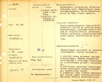
File of histopathological evaluation of nervous system diseases (1966) - nr 84/66
Kartoteka oceny histopatologicznej chorób układu nerwowego (1966) - opis nr 84/66 - Współwytwórcy:
-
K.Wiśniewska, dr
K. Wiśniewska, dr - Słowa kluczowe:
-
Choroby naczyniowe - krwotoki
Vascular diseses - hemorrages
Krwotok mózgowy, podpajęczynówkowy - Data publikacji:
- 1966
- Język:
- polski
- Linki:
- https://rcin.org.pl/dlibra/publication/edition/193364/content Link otwiera się w nowym oknie
- Prawa:
-
Creative Commons Attribution BY 4.0 license
Licencja Creative Commons Uznanie autorstwa 4.0 - Dostawca treści:
- RCIN - Repozytorium Cyfrowe Instytutów Naukowych
- Książka
Menu główne
Wyszukiwarka
Treść główna
